Patent Foramen Ovale Vs Asd In Clark
Description


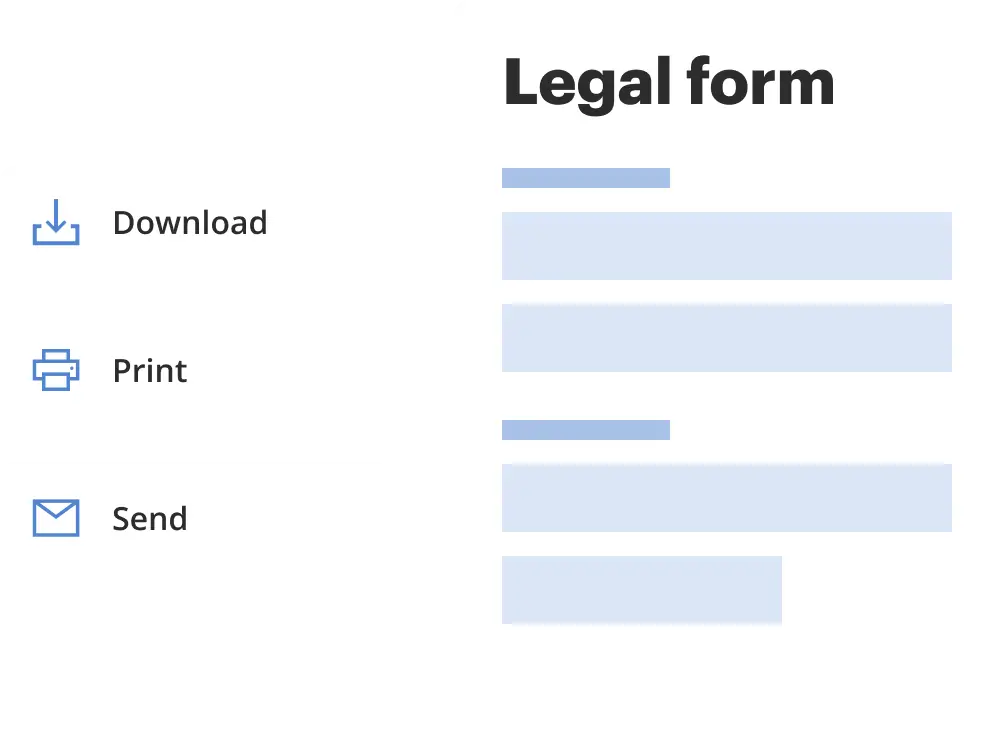
Get your form ready online
Our built-in tools help you complete, sign, share, and store your documents in one place.
Make edits, fill in missing information, and update formatting in US Legal Forms—just like you would in MS Word.
Download a copy, print it, send it by email, or mail it via USPS—whatever works best for your next step.
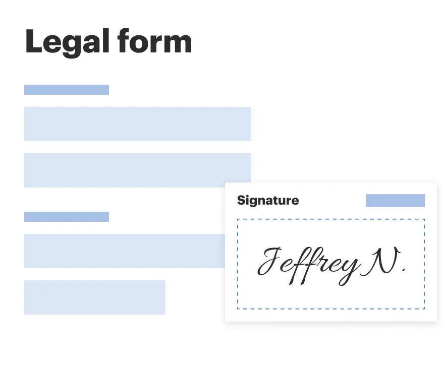
Sign and collect signatures with our SignNow integration. Send to multiple recipients, set reminders, and more. Go Premium to unlock E-Sign.
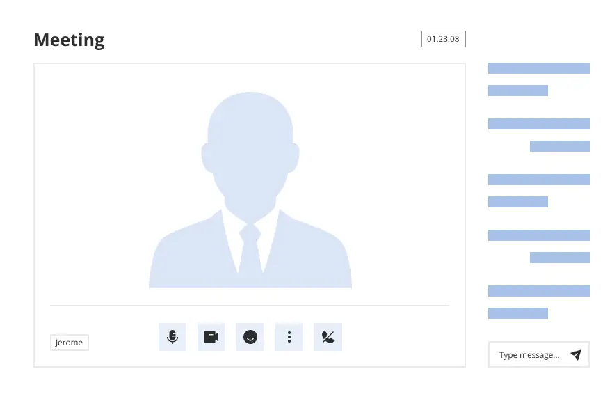
If this form requires notarization, complete it online through a secure video call—no need to meet a notary in person or wait for an appointment.
We protect your documents and personal data by following strict security and privacy standards.
Make edits, fill in missing information, and update formatting in US Legal Forms—just like you would in MS Word.
Download a copy, print it, send it by email, or mail it via USPS—whatever works best for your next step.
Sign and collect signatures with our SignNow integration. Send to multiple recipients, set reminders, and more. Go Premium to unlock E-Sign.
If this form requires notarization, complete it online through a secure video call—no need to meet a notary in person or wait for an appointment.
We protect your documents and personal data by following strict security and privacy standards.
Looking for another form?
Form popularity
FAQ
CT diagnosis of PFO was defined as (1) a channel-like appearance of the interatrial septum (IAS) and (2) a contrast agent jet flow from the left atrium (LA) to the right atrium (RA). ASD was defined as (1) the IAS resembling a membrane with a hole and (2) a contrast jet flow between the two atria.
ICD-10 code Q21. 12 for Patent foramen ovale is a medical classification as listed by WHO under the range - Congenital malformations, deformations and chromosomal abnormalities .
However, although clinically very different, atrial septal defects (ASD) and patent foramen ovale (PFO) share a single diagnostic code (ICD-9: 745.5, ICD-10: Q21. 1).
CPT® Code 33641 - Repair Procedures for Septal Defect - Codify by AAPC.
Intracardiac echocardiography (ICE) (CPT‡ code +93662) during therapeutic/diagnostic intervention, including imaging supervision and interpretation (List separately in addition to code for primary procedure).
Small holes between the upper chambers of the heart may be categorized as an atrial septal defect (ASD) or patent foramen ovale (PFO). An ASD is a congenital heart defect, a condition that you are born with. PFOs can occur only after birth when the foramen ovale fails to close.
Question: How should I report PFO/ASD closure using an ICE catheter for guidance? Idaho Subscriber Answer: For patent foramen ovale (PFO) and atrial septal defect (ASD) closure, you should assign 93580 (Percutaneous transcatheter closure of congenital interatrial communication i.e., Fontan ...
Idaho Subscriber Answer: For patent foramen ovale (PFO) and atrial septal defect (ASD) closure, you should assign 93580 (Percutaneous transcatheter closure of congenital interatrial communication i.e., Fontan ...
``In simplistic terms, a PFO is the result of incomplete closure of atrial tissue, whereas an ASD is the result of complete absence of such tissue between the right and left atrial heart chambers.''
During the procedure, an interventional cardiologist: Makes a tiny incision in your groin and inserts a catheter into a blood vessel. Uses ultrasound imaging to get a closer view of the PFO. May guide a deflated balloon through the catheter and move it to the PFO.